| UNIVERSITY OPHTHALMOLOGY CONSULTANTS |
| UNIVERSITY OPHTHALMOLOGY CONSULTANTS |
|
CASE OF THE MONTH CASE #1 |
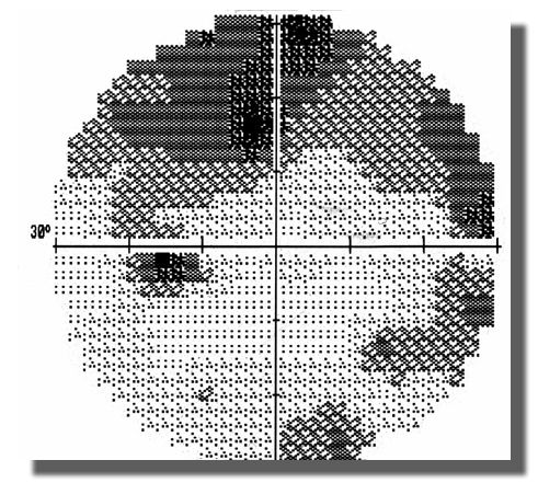
| Follow-up: The patient was uncooperative and failed to comply with both the plan we had elaborated with her primary care physician (PCP) and our own follow-up. She ignored our requests for follow-up and failed to keep appointments with her PCP. She finally returned for examination 6 weeks later, after receiving a registered letter. Visual acuity was 20/25 OS with mild color vision changes and early arcuate visual field loss in her only eye. The patient had continued to experience severe headaches. |
| FIGURE 9. Image of the visual field 6 weeks after initial neuro-ophthalmic evaluation (previously normal) |
 |
| Hospital admission: Arrangements were made with the patient’s PCP, and the patient was admitted to the hospital. Three days later, visual acuity was 20/200 OS. The patient identified correctly only 2 of 10 color plates, evidence of marked visual field loss with increased disc edema.The x-ray film of the chest showed no abnormalities. We performed a lumbar puncture, which revealed a protein content of 180 mg/dL and a cell count of 210 cells/cu mm, 97% of which were lymphocytes. |
| WHAT IS YOUR DIAGNOSIS AND SUGGESTED TREATMENT MODALITY? |
| Click here for outcome |