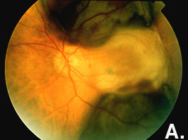
Figure A. Retinal photograph obtained when the photographic
session precedes the echographic examination. Note the clarity of
the media.
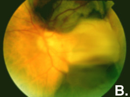
Figure B. Retinal photograph obtained when the
photographic session follows the echographic examination. The image’s
low quality prevents true color rendition of the retinal structures.
EXPLANATION:
During echography, the topical application of proparacaine hydrochloride
and methylcellulose to the corneal surface causes temporary swelling
of the corneal epithelial cells. This effect lasts for at least 1
hour and persists even when the cornea is carefully rinsed.

