| FUNDUS PHOTOGRAPH | FLUORESCEIN ANGIOGRAM |
 |
 |
|
|
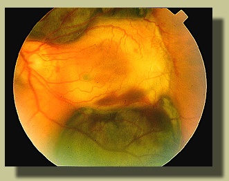
The fundus appearance OS is due to disciform scar with adjacent subretinal hemorrhage at its superior and inferior borders. The blood appears green-brown. No RPE-level detail (e.g., drusen) is evident due to the subretinal location of the blood. |
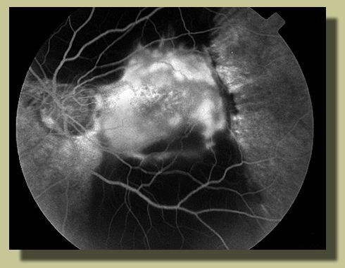
Fluorescein angiogram OS reveals normal retinal vasculature and blockage of underlying choroidal fluorescence due to subretinal blood *. | |
| DIFFERENTIAL DIAGNOSIS: This case illustrates subretinal hemorrhage mimicking choroidal melanoma. | ||
| * |
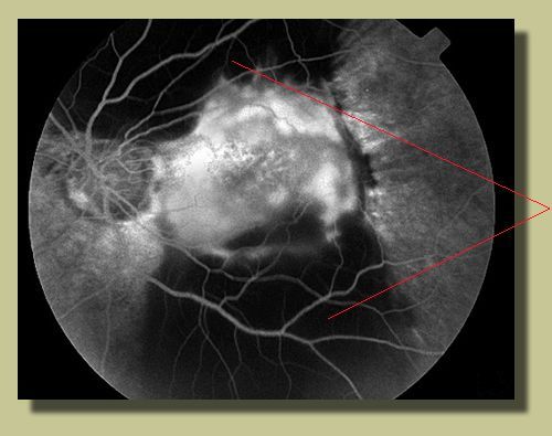 |
The
blockage of underlying choroidal fluorescence on the fluorescein angiogram
is due to subretinal blood.
|
| Please click here to return to case presentation |
| Please send comments to: Dr. Marco Zarbin at zarbin@umdnj.edu |
| This case appeared in the following article: |
| Hochman MA, Seery CM, and Zarbin MA. Pathophysiology and management of subretinal hemorrhage. Surv Ophthalmol 1997; 42(3):195-213. |
| Reprinted with permission of Survey of Ophthalmology |
| residency program /// patient care services /// research | |
| ophthalmic medical assistant program /// continuing medical education | |
| facilities /// faculty /// library /// faqs | |
|
This site is under development by SciMedWeb