|
|
|
Case #8
|
Typical Clinical Presentation:
|
An incidental finding of a bone lesion in the distal tibial meta-diaphysis of an 13-year-old male.
The lesion was totally asymptomatic.
-
As always, pay attention to the patient's age. The fact that the lesion did not produce symptoms
and was found incidentally suggests benignancy.
|
|
|
|
Characteristic Radiological Findings:
|
|
|
|
-
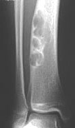
Plain radiograph shows a sharply demarcated, lucent, loculated, meta-diaphyseal lesion
surrounded by a rim of sclerotic bone. The lesion predominantly involves the lateral portion
of the bone and produces mild cortical expansion.
|
|
Radiological findings are so typical that the diagnosis can be made with certainty by X-ray alone provided the
lesion is in the typical skeletal site and appropriate age group. Note the eccentric location along the long axis of the bone.
Large lesions can involve the entire diameter of the bone expanding the cortex. However, they are always sharply
demarcated and rimmed by sclerotic bone.
|
|
|
|
Pathological Findings:
|
|
|
|
-
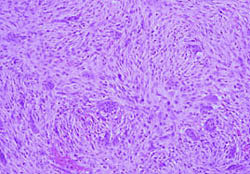
Curettage specimen consisted of firm red-tan tissue with few small yellowish areas.
Low power view showed a moderately cellular lesion composed of uniform spindle cells
in a storiform pattern and scattered giant cells; .
|
|
|
|
-
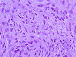
Spindle cells had bland appearance. Mitotic figures were easily found averaging 4 per 10 hpf.
However, no atypical mitoses were identified.
|
|
|
|
-
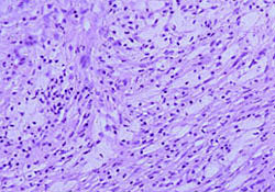
Multiple collections of foamy histiocytes (xanthoma cells) were seen throughout the lesion.
Other fields showed hemosiderin-laden macrophages.
|
|
|
Diagnosis: Non-Ossifying Fibroma (NOF)
(fibrous cortical defect, or metaphyseal cortical defect)
|
|
Salient Points::
|
-
NOF is a common, non-neoplastic, self-healing lesion occuring in skeletally immature individuals, usually between
the ages of 5 and 20 years. Small lesions are usually incidental radiological findings. The larger lesions occupying more
than a half of the bone diameter may present with a pathologic fracture.
-
Location. In most cases, NOF presents as a solitary lesion in the metaphysis or meta-diaphysis of the long bone
at the knee (distal femur, proximal tibia or fibula), distal tibia and proximal humerus. A syndrome of multiple
non-ossifying fibromas and cutaneous cafe au lait spots is known as Jaffe-Campanacci syndrome.
-
For lesions that have the histologic features of NOF but occur in unusual locations such as pelvis, ribs or vertebrae,
some authors use the term "benign fibrous histiocytoma". However, the designation remains controversial and is not
generally accepted.
-
GCT may enter your differential diagnosis. Remember, however, that it is characterized by the epiphyseal
location and occurrence in adults.
Available publications for the topic:
Non-Ossifying Fibroma
|

