| UNIVERSITY OPHTHALMOLOGY CONSULTANTS |
| UNIVERSITY OPHTHALMOLOGY CONSULTANTS |
|
CASE OF THE MONTH CASE #25
|
| FLUORESCEIN
ANGIOGRAM SERIES OS (July 27, 2001) |
 |
 |
|
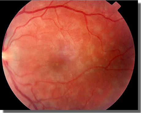
FUNDUS IMAGE OS. Macula
with whitish lesions at the |
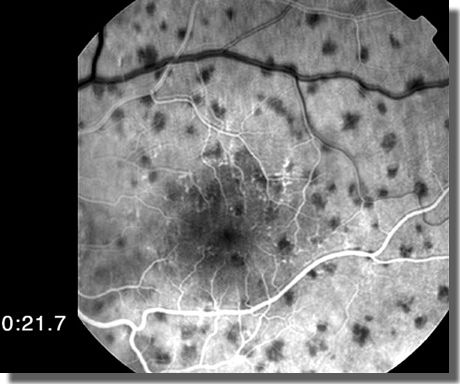
EARLY PHASE ANGIOGRAM. Choroidal perfusion defects and an early wreath-like pattern of hyperfluorescence correspond almost uniformly with areas of RPE-level whitening seen on the fundus photograph on the left. |
|
|
|
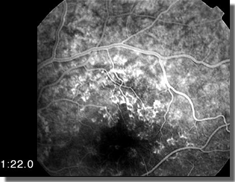
LATE VENOUS FILLING PHASE.
Areas of RPE-level whitening demonstrate a wreath-like pattern of hyperfluorescence
at the perimeter |
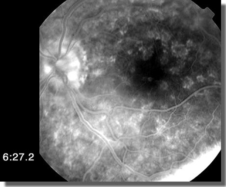
RECIRCULATION PHASE. Hyperfluorescence and leakage of dye from the optic nerve head. |