|
The patient became cushingoid due to steroid use. Because
of the indeterminate pathological findings, the improvement in
the patient’s clinical condition, and the possibility
that MEWDS or a related condition was the cause of uveitis, oral
steroid treatment was tapered and the patient was followed-up
for recurrent inflammation.
4 WEEKS AFTER
STOPPING PREDNISONE: Areas of RPE depigmentation
were noted in the inferotemporal midperiphery.
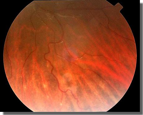
|
Color photograph demonstrating areas of RPE depigmentation
and perivascular sheathing of large choroidal vessels in
the mid periphery of the left fundus
(April 12, 2002).
|
 |
6
WEEKS AFTER STOPPING PREDNISONE: Once again, the patient
reported decreased vision. Examination was
notable for the following:
- Visual acuity of 20/20- OS,
- 1-2+ anterior chamber cell and flare,
- 1+ vitreous cell, and
- Swelling of the optic nerve head.
- A repeat fluorescein angiogram showed areas of blocked choroidal
fluorescence early, but not to the extent that had been seen
before, and late leakage. Mild leakage from the optic nerve
head was present. Periphlebitis was evident for the first time.
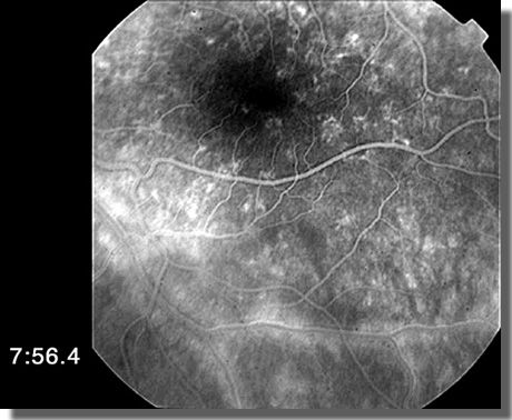
|
Late phase fluorescein angiogram demonstrating dye leakage
associated with periphlebitis in the left eye
(April 23, 2002).
|
 |
- A repeat visual field revealed a normal-size blind spot.
|