| UNIVERSITY OPHTHALMOLOGY CONSULTANTS |
| UNIVERSITY OPHTHALMOLOGY CONSULTANTS |
|
CASE OF THE MONTH CASE #1 |
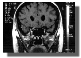
| FIGURES 4-6: Gadolinium-enhanced T1 coronal view of the optic chiasm/posterior optic nerves |
| FIGURE 4 |
 |
| Click for interpretation of MRI scans |