| External Eye
Examination |
|
EXTERNAL
VIEW OD |
| |
|
| |
 |
| |
............................................................. |
| |
No
lid edema OU |
| |
|
|
Visual Acuity |
OD:
20/400
OS: 20/20
• No improvement to pinhole OU |
| |
|
| Extraocular
Motility |
Full
(pupils were previously dilated by ophthalmologist) |
| |
|
| Pupil
Examination |
Pupils
were dilated by non-affiliated ophthalmologist |
| |
|
| Intraocular
pressure with applanation tonometry |
OU:
20 mm Hg |
| |
|
| Confrontation
Visual Fields |
Full
to finger counting OU |
| |
|
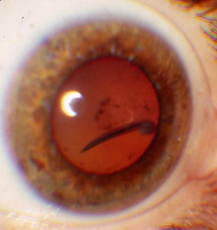
| Slit
Lamp Examination |
OD
• Normal eyelids, lacrimal glands, and lashes
• Trace temporal conjunctival hyperemia
• Clear and smooth cornea
• 1-2+ cell and flare in the anterior chamber
• Trace pigment on anterior capsule
• Clear lens
• 2-3+ vitreous cells
• Presence of a nematode, measuring approximately 0.5
x 4 mm, in the anterior vitreous cavity
OS: Unremarkable
|
| |
|
| Fundus
Examination |
OD
• 2-3+ vitritis was noted
• An intraocular parasite with columnated blood, reddish,
approximately 0.5 x 4 mm in size, was noted floating in
the anterior vitreous.
• The optic nerve was pink and sharp with a cup-to-disc
ratio
of 0.2.
• Presence of a 1.5 DD area of preretinal/retinal fibrosis
involving the maculo-papillary bundle. Retinal striae are
visible due to vitreous traction in this area.
• An area of chorio-retinal atrophy measuring about
3/4 DD was noted temporally along the superior arcade.
OS
• Clear vitreous
• The disc was pink and sharp with a cup-to-disc ratio
of 0.2.
• Normal fundus
|