| UNIVERSITY OPHTHALMOLOGY CONSULTANTS |
|
CASE OF THE MONTH CASE #3 |
| UNIVERSITY OPHTHALMOLOGY CONSULTANTS |
|
CASE OF THE MONTH CASE #3 |
| WHAT TESTS WOULD YOU ORDER? |
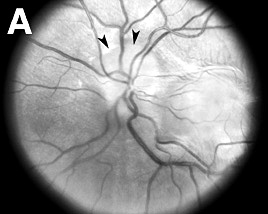
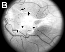
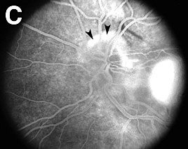
| Lumbar puncture and MRI of the head would demonstrate diagnosis of pseudotumor cerebri.Fluorescein angiography (FA) (Figure 2C) would demonstrate diagnosis of bilateral dye leakage from optic nerve head, suggestive of bilateral papilledema or bilateral papillitis, as well as vascular nature of subretinal scar OS. |
|
FIGURE
1: PATHOLOGY OS
|
| RED-FREE PHOTOGRAPHY | RED-FREE PHOTOGRAPHY | FLUORESCEIN ANGIOGRAPHY | ||||||
 |
 |
 |
||||||
|
The ophthalmologist noted a peripapillary subretinal fibrotic mass involving the left fovea, possibly due to a choroidal inflammatory lesion. Red-free fundus photography and fluorescein angiography disclosed a subfoveal CNV associated with mild swelling and late staining of the optic nerve (Figure 1). The patient was treated with a posterior subtenon prednisolone acetate (40 mg) injection. There was no improvement. |
|
| Previous page | Next page | ||