| UNIVERSITY OPHTHALMOLOGY CONSULTANTS |
|
CASE OF THE MONTH CASE #4 |
| UNIVERSITY OPHTHALMOLOGY CONSULTANTS |
|
CASE OF THE MONTH CASE #4 |
|
FIGURE
3
|
FIGURE 4 | |
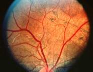
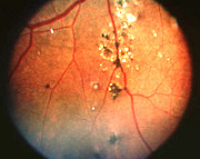
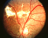
| FUNDUS PHOTOGRAPHY OD | FUNDUS PHOTOGRAPHY OD | ||||
 |
 |
||||
|
FIGURE
5
|
|
| FUNDUS PHOTOGRAPHY OS | What are the fundus findings? | |||
 |
Angioid streaks OU, subretinal fibrosis superior to the optic nerve head OD, and focal RPE hyperplasia and white subretinal crystalline bodies inferior to the inferotemporal arcade OD. |
|||
|
|
||
| Previous page | Next page | ||