| UNIVERSITY OPHTHALMOLOGY CONSULTANTS |
|
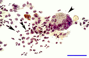
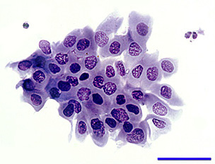
CASE OF THE MONTH CASE #6 |
|
|
| UNIVERSITY OPHTHALMOLOGY CONSULTANTS |
|
CASE OF THE MONTH CASE #6 |
|
|
| Previous page | Next page | ||