|
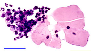
Cytopathology of the material obtained by paracentesis disclosed
polymorphonuclear leukocytes,
histiocytes, multinucleated
giant cells, and plasma
cells surrounding amorphous lens material (Figures
2 & 3).
 |
|
Figure 2. Phagocytosis of lens material
by inflammatory infiltrate comprising polymorphonuclear
leukocytes (PMNs), lymphocytes, and plasma cells.
Mag. bar = 50 mm, Romanovsky stain.
|
 |
|
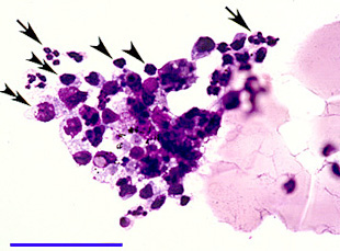
Figure 3. High magnification of Figure
2 showing PMNs (arrow), lymphocytes (arrowhead), and plasma
cells (double arrowhead).
Mag. bar = 50 mm, Romanovsky stain.
|
|