|
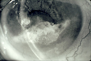
Slit lamp examination
of the left eye revealed mild conjunctival infection, a
clear cornea, and a flat anterior chamber inferiorly with peripheral
anterior synechiae
extending inferiorly from the 3:30-o’clock to the 8:30-o’clock
position. A partially opaque, white material that seemed gelatinous
and crystalline had settled inferiorly, giving the appearance
of a pseudohypopyon
(Figure 1).
| |
|
FIGURE
1 |
|
|
| |
|
 |
|
Slit lamp photograph
showing white plaque of material in the inferior anterior
chamber and posterior synechiae involving the superior half
of the pupil. |
| |
|
|
|
|
A few mutton-fat keratic precipitates on the inferior corneal
endothelium, 1+ cell, and scattered posterior
synechiae (limiting the pupillary dilation to
3 mm) were present as well. The limited view of the lens revealed
a white cataract
with no visualization of the posterior lens surface or the anterior
vitreous.
|