| WHAT
ARE THE ECHOGRAPHIC FINDINGS? |
|
Elongated globe. Phakic eye. The vitreous cavity presents medium
reflective, mobile, point- and membrane-like echoes. A highly reflective
retinal membrane with limited aftermovement is seen in the inferotemporal
quadrant extending between the 5:30-o’clock and the 7:30-o’clock
meridian at the equator, with its highest elevation at the 7:30-o’clock
position. This membrane does not present any retinal tears or traction.
Behind it are low reflective, mobile, point-like echoes with adjacent
thickening of the choroid. Macula presents cystic thickening of
the retinal-choroidal layer. No scleral rupture is observed. The
optic nerve head is found in the posterior scleral depression. The
orbital fat pattern is within normal limits.

DIAGNOSTIC IMPRESSION
OD
- 1. Posterior staphyloma
- 2. Phakic globe
- 3. Vitreous hemorrhage
- 4. Inferotemporal retinal detachment
- 5. Macular edema vs hemorrhage
- 6. Diffuse choroidal thickening of the
posterior pole (hemorrhage)
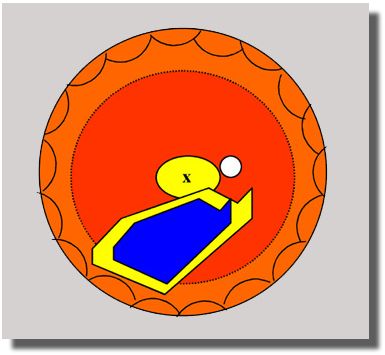 |
YELLOW:
Areas of thickening at the macula
and choroid adjacent to the retinal detachment
BLUE:
Retinal detachment
|
|
|