| UNIVERSITY OPHTHALMOLOGY CONSULTANTS |
| UNIVERSITY OPHTHALMOLOGY CONSULTANTS |
|
CASE OF THE MONTH CASE #25
|
| FUNDUS PHOTOGRAPHS OS |
 |
 |
|
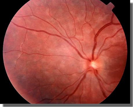
A. Fundus photograph of left optic nerve head and nasal periphery
demonstrates mild blurring of the nerve head margin and, nasally, |
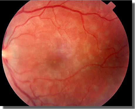
B. Fundus photograph of the left macula. Whitish lesions at the level of the RPE range from 300 to 500 µm in diameter. |