| UNIVERSITY OPHTHALMOLOGY CONSULTANTS |
|
CASE OF THE MONTH CASE #3 |
|
FIGURE
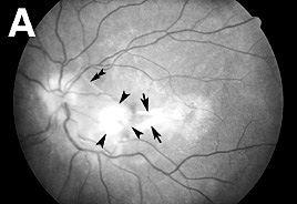
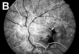
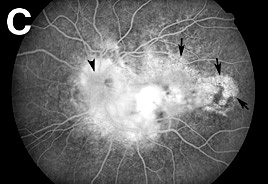
2: PATHOLOGY OS
|
| 5 YEARS LATER |
| RED-FREE PHOTOGRAPHY | FLUORESCEIN ANGIOGRAPHY | FLUORESCEIN ANGIOGRAPHY | ||||||
 |
 |
 |
||||||
|
| The fundus examination was significant for bilateral papilledema with moderate optic nerve pallor OS (Figure 2A). In addition, a 1 disc diameter subretinal CNV with subfoveal extension and associated exudative retinal detachment was present in the left eye. The overlying retina showed prominent parafoveal cysts. The FA disclosed early hyperfluorescence of the scar with window defects temporally where there was RPE atrophy (Figure 2B-C). |
|
| Previous page | Next page | ||