| UNIVERSITY OPHTHALMOLOGY CONSULTANTS |
|
CASE OF THE MONTH CASE #5 |
|
FIGURE
2: PREOPERATIVE FINDINGS
|
| FUNDUS PHOTOGRAPHY OS |
| FIGURE 2A | FIGURE 2B | FIGURE 2C | ||||||
 |
 |
 |
||||||
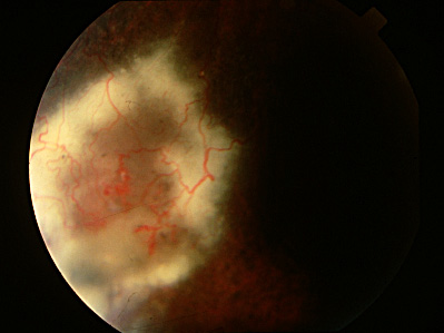
| 2A. Normal-appearing left optic nerve head and macula. 2B. Area of retinal telangiectasia with surrounding lipid in the inferonasal periphery of the left fundus. 2C. Inferotemporal area of retinal telangiectasia OS before laser photocoagulation. | ||||||||