| FINDINGS:
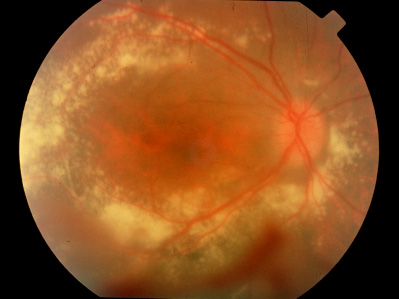
Pink discs with sharp margins, a cup-to-disc ratio of
0.0, and attenuated arterial caliber at the optic nerve head OU. Intraretinal
and subretinal lipid deposits were present in the area centralis OD. RPE
hyperplasia was noted in the midperiphery for 360 degrees OU. On the right,
2 areas of telangiectasia were present. One, centered just anterior to
the equator at the 4:30-o’clock position, was associated with intraretinal
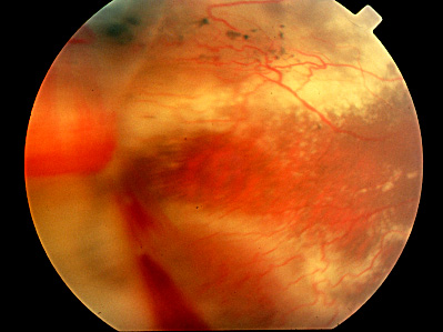
lipid and very mild exudative retinal detachment. Another area of retinal
telangiectasia was present midway between the equator and the ora serrata
and extended from the 8-o’clock to the 10-o’clock position.
Near the anterior margin of the telangiectatic lesion, a frond of retinal
neovascularization with associated vitreous hemorrhage, extended from
the 8:30-o’clock to the 10-o’clock position. Vitreoretinal traction
in this location created a retinal detachment from the 8:30-o’clock
to the 9:30-o’clock position, with the posterior margin 5 disc diameters
temporal to the fovea, with subretinal fibrosis. In addition to bone spicule
RPE hyperplasia, areas of placoid RPE hyperplasia and RPE atrophy were
present in the midperiphery of attached retina. On the left, an area of
retinal telangiectasia was present at the equator and was centered on
the 6:30-o'clock meridian (not shown). The lesion was surrounded by intraretinal
lipid and exhibited localized, underlying mild exudative retinal detachment
and overlying mild fibrosis in the vitreous. |