|
 VITRECTOMY:
The CNV was excised via a conventional 3-port
vitrectomy with subretinal
dissection. The posterior two thirds of the vitreous gel including the
posterior vitreous cortex was excised. The preoperative
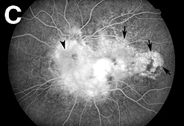
FA (Figure 2C) was projected in the operating room to
guide the subretinal dissection. There was a substantial exudative retinal
detachment and prominent cystic change not just in the parafoveal area,
but even outside the foveal avascular zone. The retina was puckered
in an area of foveal adherence to the underlying scar.
The subretinal fibrosis extended from the temporal margin of
the optic nerve to the fovea. VITRECTOMY:
The CNV was excised via a conventional 3-port
vitrectomy with subretinal
dissection. The posterior two thirds of the vitreous gel including the
posterior vitreous cortex was excised. The preoperative
FA (Figure 2C) was projected in the operating room to
guide the subretinal dissection. There was a substantial exudative retinal
detachment and prominent cystic change not just in the parafoveal area,
but even outside the foveal avascular zone. The retina was puckered
in an area of foveal adherence to the underlying scar.
The subretinal fibrosis extended from the temporal margin of
the optic nerve to the fovea.
|