| UNIVERSITY OPHTHALMOLOGY CONSULTANTS |
|
CASE OF THE MONTH CASE #3 |
| UNIVERSITY OPHTHALMOLOGY CONSULTANTS |
|
CASE OF THE MONTH CASE #3 |
| HISTOPATHOLOGY OF EXCISED CNV |
| FIGURE 3A | FIGURE 3B | |||
 |
 |
|||
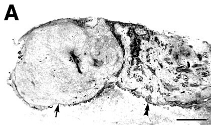
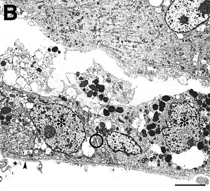
| Histologically, the excised CNV disclosed a specimen consisting of a vascular component and a collagenous scar (Figure 3A). Hyperplastic RPE lined the surface of the scar, and probable RPE was present in the interior of the scar. Electron microscopy showed discontinuous basement membrane lining the surface of the scar (Figure 3B). | |
| FIGURE 3C | FIGURE 3A | |||
 |
 |
|||
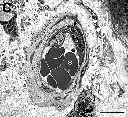
| The vascular component of the CNV contained well-defined vascular channels centrally (Figure 3C) and was surrounded by RPE (Figure 3A). | ||||
| FIGURE 3D | FIGURE 3E | |||
 |
 |
|||
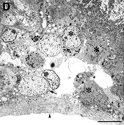
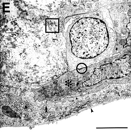
| Clumps of hyperplastic RPE, basement membrane (Figures 3D and E), and widely spaced collagen (Figure 3E) were also present. Neither the collagenous components of Bruch’s membrane nor the choriocapillaris were noted in the specimen. | |
|
| Previous page | Next page | ||