| UNIVERSITY OPHTHALMOLOGY CONSULTANTS |
|
CASE OF THE MONTH CASE #3 |
| UNIVERSITY OPHTHALMOLOGY CONSULTANTS |
|
CASE OF THE MONTH CASE #3 |
| OUTCOME |
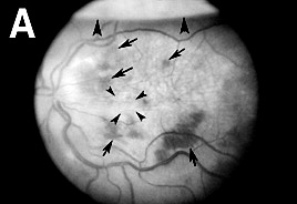
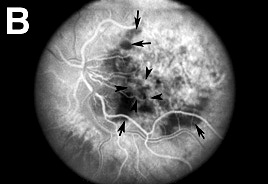
| FIGURE 4: ONE WEEK AFTER SURGERY |
| RED-FREE PHOTOGRAPHY | FLUORESCEIN ANGIOGRAPHY | ||||
 |
 |
| One week after surgery, an area of atrophy or hypopigmentation at the level of the RPE was evident in the previous location of the CNV (Figure 4A). Fluorescein angiography showed mottled subfoveal choriocapillaris fluorescence (Figure 4B). |
|
| Previous page | Next page | ||